Biology · molecular structure
DNA, RNA and genetic information
A cut on your skin can heal because cells make new material and, in the right circumstances, divide. But how does a new cell receive the information it needs? And how does a cell use stored information to make something that actually works? These are different questions. DNA copying answers the first; RNA and protein production help answer the second.
If DNA, genes and RNA already sound like a list of names to remember, leave that list aside for a moment. We have two concrete questions: how information is passed on, and how it is used. We will keep those questions beside us as we move from a cell to one selected piece of DNA. A gene, a nucleotide and a ribosome will each get an explanation when it has a job in our example; their meanings will remain beside the model.
Our main setting is a human cell with a nucleus. The molecule shapes, spacing, colours and teaching sequences are simplified; they are not a time-resolved recording of a living cell or an atomic structure measurement.
1. Start with a cell that has work to do
Your body contains many cells. A cell is a small living unit. A membrane separates its inside from its surroundings and controls what crosses it; it is not a solid wall that nothing can pass through. Inside, molecules take part in reactions that make new material or release usable energy, for example by breaking down sugar. Other molecules help move substances around. A molecule is a group of connected atoms; atoms are very small building units of matter, and a chemical reaction rearranges them into different combinations. For this chapter, keep three things in view: a cell, its membrane, and the molecules that do its work.
Many of the molecules doing this work are proteins. A protein contains one or more chains of amino acids, which are small molecular building units. The order of those units affects how the chain folds and what the protein can do. We therefore need more than a supply of raw material: the cell also needs a way to specify useful arrangements of that material.
The order we need to preserve brings us to DNA, whose full name is deoxyribonucleic acid. It is a chemical substance that stores biological sequence information. Calling it information can feel abstract for now. Later we will follow the order of its units and see how other molecules use that order; for the moment, let us locate the material itself. Most DNA in the human cell model is in the nucleus, a compartment with its own surrounding membrane. A small amount also occurs in mitochondria; our example follows the nuclear DNA.
2. Follow the same material, without losing where it came from
Enlarging a tiny structure can create a new difficulty: we can see more detail, but lose track of where it came from. A cell and a DNA molecule differ enormously in size, so we need several changes of magnification. Find the orange selection before pressing Next. That colour keeps hold of the same selected material while the view changes. If the enlarged view becomes unfamiliar, glance at the small context drawing; it keeps the earlier location in view for us.
The purple threads inside the nucleus represent chromatin. Chromatin means DNA together with proteins associated with its organisation. The orange section is a selected part of those same purple threads. When we later distinguish DNA from the proteins around which it is wrapped, we are revealing the composition of chromatin, not finding a second, unrelated material concealed inside it.
Purple = chromatin. Orange = one selected part of that same chromatin.
A cell and its nucleus
We begin with a simplified animal cell. The outer green boundary is the cell membrane. Inside it, the smaller purple compartment is the nucleus. We have removed the front of both boundaries so that you can see inside; a real cell does not have these open faces.
Find the orange-marked part inside the purple nucleus. We will keep following that same selected part.
Look back at the wrapping unit. Its name is a nucleosome: DNA wrapped around a core of eight histone proteins. A histone is a kind of protein, not another name for DNA. Many such units help organise a long DNA molecule. The model spreads units apart so that their connections remain visible; chromatin in a cell is not always a uniform, regularly coiled fibre.
DNA and chromosome are also not interchangeable names. A chromosome is an organised unit of this DNA–protein material. Before DNA is copied, one human nuclear chromosome contains one long double-stranded DNA molecule with associated proteins. It remains a chromosome when it is spread out and difficult to distinguish under a microscope; it does not have to look like an X.
3. Why an X contains two DNA molecules
For counting, take a typical human body cell with two sets of chromosomes: 23 from each parent, giving 46 in total. This is our example, not a claim about every human cell. Egg and sperm cells normally have 23; mature human red blood cells have no nucleus. Some other specialised cells also differ from this example.
Before an ordinary cell division, each DNA molecule is copied. The two copies remain associated in two sister chromatids. A chromatid is one of these DNA-and-protein copies. The sisters are held together until they separate; the constricted centromere region is the familiar joining region in an X-shaped drawing. Each sister contains a complete double-stranded DNA molecule. The two sides of the X are therefore not the two strands of one DNA double helix.
While the centromere holds the sisters together, they are one object, so we still count the pair as one replicated chromosome. DNA amount has doubled, but the chromosome count has not yet doubled. Once the sisters separate, each is counted as a chromosome in its own right. In ordinary division, the two daughter cells each receive one copy of every chromosome.
Follow one chromosome
1 chromosome · 1 DNA molecule
The thick line is one DNA–protein unit. It represents an entire chromosome schematically, not one strand of a DNA molecule.
The sketch above follows one chromosome. To see all of them, return to the 3D model in chapter 2 and press “Compare the dividing-cell chromosomes”: that division view shows 46 replicated chromosomes, containing 92 DNA molecules. It arranges them with space between them so that you can inspect the count; it is not their random live arrangement or a metaphase plate. Their X-like shape represents condensed replicated chromosomes, not the special identity of the X sex chromosome. Nuclear-envelope breakdown and chromosome condensation occur over stages, not as one instantaneous switch.
4. Build a strand, then pair it with another
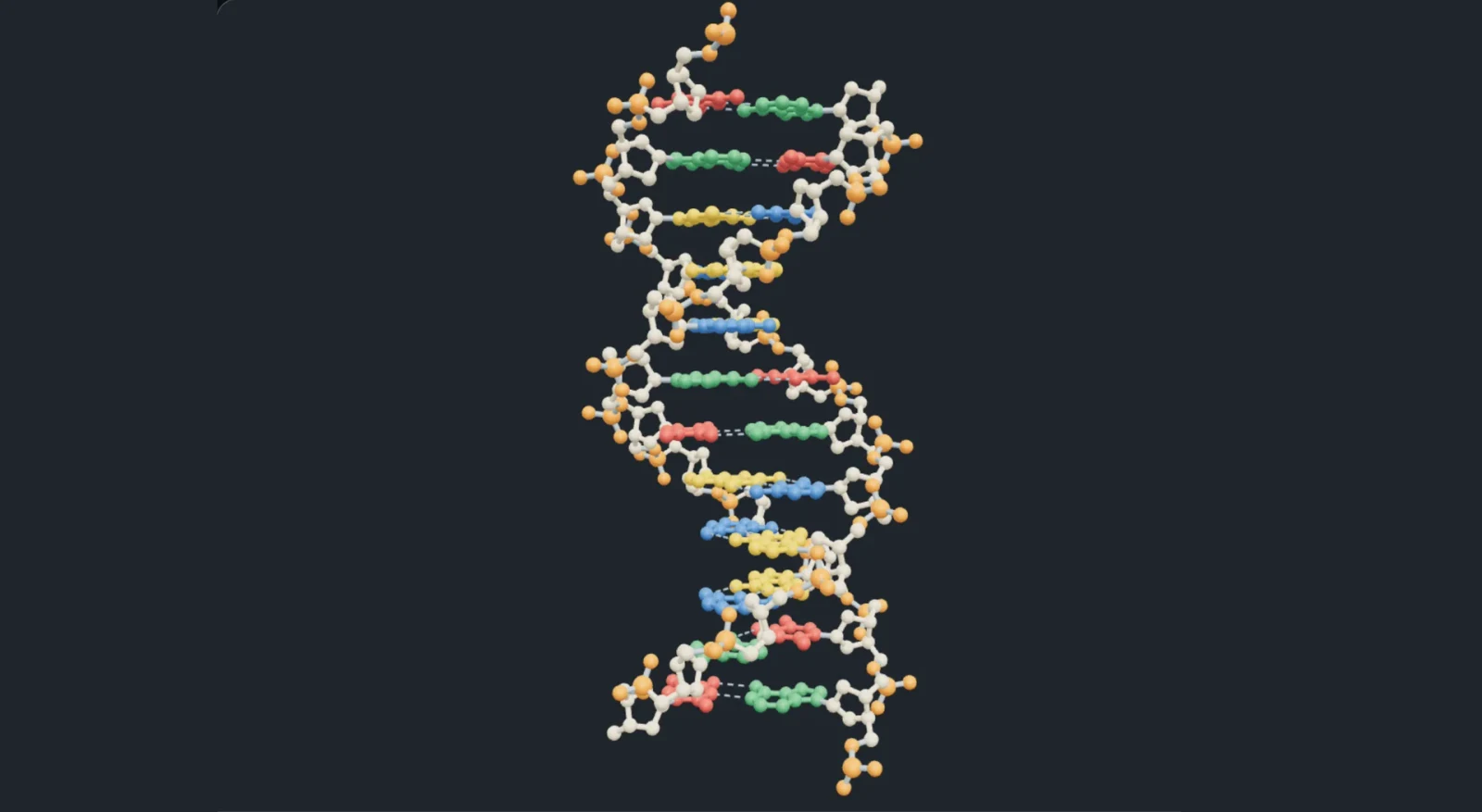
We now return to the DNA molecule itself. Matter is made from atoms, and chemical bonds can join atoms into molecules. A nucleotide is one repeating unit of a DNA strand. It has three connected parts: a phosphate group, a sugar called deoxyribose, and a base. Here “base” names one part of a nucleotide; it does not mean the whole nucleotide.
DNA uses four usual bases: adenine (A), thymine (T), guanine (G) and cytosine (C). We use the letters because it is easier to follow a sequence of twelve letters than twelve long chemical names. Repeated sugar–phosphate connections make the backbone of a strand; each sugar also carries its base. Changing the letter changes the base, not the basic arrangement of the entire backbone.
Put a second strand beside the first. In the usual DNA pairing, A pairs with T, and G pairs with C. These complementary pairs have compatible shapes and arrangements of atoms that can form hydrogen bonds. Hydrogen bonds are attractions between particular groups of atoms. They differ from the covalent bonds that hold each strand’s backbone together.
One nucleotide
A nucleotide is one building unit of a DNA strand. Orange marks its phosphate group; cream marks its sugar, called deoxyribose; green marks the base A. The sticks represent covalent bonds joining atoms. Select a part to make the other parts quieter, then reset the view or drag to see that this is a solid three-dimensional structure.
The balls are a drawing convention for atoms, not their literal surfaces. Most hydrogen atoms are omitted to keep the bonds readable.
A–T normally forms two hydrogen bonds and G–C three. But it would be incomplete to say that hydrogen bonds alone hold DNA in its familiar form. Neighbouring bases also stack against one another inside the helix, and these stacking interactions contribute substantially to its stability. In the model, inspect both across a pair and along the stack.
Some parts of a molecule really are approximately flat: the rings of these bases are an example. Other parts extend in three dimensions. A faithful model should retain both facts. Drag the nucleotide to inspect its puckered sugar and spatial phosphate group, then compare that with a base ring. The balls and sticks are a convention for atoms and bonds, not a photograph of their surfaces.
5. A sequence needs a direction
The sugar in each nucleotide has numbered carbon atoms. The labels and refer to two of those positions, read “five-prime” and “three-prime”. Successive nucleotides connect through a phosphate linking the position of one sugar to the position of the next. The resulting strand has two chemically different ends. The prime mark distinguishes sugar-atom numbering; it is not a derivative, a unit of time or a length.
DNA’s two strands run in opposite directions. If the upper strand runs from on the left to on the right, the lower partner runs from on the left to on the right. This is called antiparallel. It describes the chemical orientation of two strands, not a claim that the strands are moving in opposite directions.
Use the same twelve positions as the structure model. At each position, choose the partner directly underneath the existing base. Start with position 1: the upper base is , so the lower one must be . At position 2 the upper base is , so the lower one must be . We are recording paired positions from left to right; we are not yet rewriting the lower strand in its own reading order.
Build the complementary strand, one position at a time
Select a numbered position to change its base using A, T, G or C below. The highlighted column contains the two bases facing one another.
At position 1, what pairs with A?
0 of 12 partner positions filled. Both strands keep their own chemical direction.
When a DNA sequence is written on its own, it is usually written from to . Finding the complementary strand means choosing the matching base at every position. That gives us the bases, but their displayed order may run from to . To write this strand in -to- order, we reverse the displayed order when needed. For these twelve positions, the lower row reads 3′–TACGCATGCGAT–5′ from left to right. Read from position 12 back to position 1, it is 5′–TAGCGTACGCAT–3′. This changes the way we write the sequence, not the molecules themselves.
6. What does it mean for DNA to contain information?
The sugar–phosphate framework repeats, but the order of bases can vary. ATG and AGT contain the same three letters in different orders. Molecules that interact with these stretches can distinguish that order. Thus a sequence can affect what a cell makes and when it makes it. Information here means an arrangement that can be copied and used through molecular interactions; the letters do not know what they mean.
A gene is a DNA sequence used to make an RNA molecule with a role in the cell. Some of these RNA molecules carry the instructions for a protein: the cell reads the relevant part of the RNA to choose the amino acids. Other RNA molecules do their work without being used to make a protein. We will meet two examples, tRNA and rRNA, shortly. So “every gene makes a protein” would leave out an important group of genes.
Nor is every part of DNA a protein-coding sequence. Some regions help regulate when genes are used; other regions have structural or other roles, and not every region’s role is fully understood. The complete collection of an organism’s DNA is its genome. A chromosome is one organised part of that collection, not one gene; many genes and other sequences can lie along it.
The twelve-base example above was invented to practise structure and pairing. It is not presented as an entire working human gene. Having the letters A, T, G and C, or even a short familiar sequence, is not enough by itself to specify a complete gene with all the signals needed for its use.
How can four letters store so many different sequences?
One position has four choices: A, T, G or C. Now add a second position. For every first choice there are four second choices: after A we can write AA, AT, AG or AC, and the same four choices follow each of T, G and C. There are therefore four groups of four, giving 16 two-letter sequences.
AA AT AG ACTA TT TG TCGA GT GG GCCA CT CG CCA third position gives four extensions of every two-letter sequence, so the count becomes . Each further position multiplies the count by four again. Writing the repeated multiplication as a power, positions allow sequences. Here counts positions, not chromosomes or amino acids.
A T G C4 choicesA T G C4 choicesA T G C4 choices7. Copy the DNA while keeping track of the old strands
Return to our first question: how can two daughter cells receive the DNA information? Simply cutting one molecule in half would give each cell only part of its sequence. Instead, before division, the cell builds a copy. Replication is the name for this copying of DNA into DNA.
Complementary pairing provides a way to do this. Separate the two strands without cutting either backbone. Each exposed old base now constrains which new base should be placed beside it. An old A calls for a new T; an old T calls for a new A; G and C similarly choose one another. The old strand serves as a template: a pre-existing sequence used to determine the new sequence.
In the model, purple backbones mark material that already existed and blue backbones mark newly made material. Base colours still identify A, T, G or C. Follow both old strands all the way to the end. Do not let “two new DNA molecules” make you assume that every atom in both products must be new.
Old5′ ATGCGTACGCTA 3′
New3′ ············ 5′
Old3′ TACGCATGCGAT 5′
New5′ ············ 3′
In the initial 3D view, positions run from bottom to top; in these letter rows, the same positions run from left to right. Dots are not yet filled. The rows compare partners; they do not show synthesis direction.
Playback holds each stage for nine seconds. Pause or use Next whenever you want more time.
Start with one double-stranded piece
Before a cell divides, it must copy its DNA. We follow only twelve base pairs, not a whole chromosome. Both backbones are purple here because both are already present. Base colours still mean A, T, G and C. The rows below record the same positions in every stage.
One DNA molecule has two strands. Keep those two meanings separate as the number of molecules changes.
At the end, each double helix contains one old strand and one new strand. That is semiconservative replication: half of each product’s two-strand framework is retained from the original molecule. The old strand supplies the sequence pattern; free nucleotides supply new material. The old bases do not split into duplicate bases or turn into the new strand.
Real cells need more than a pairing rule. This paragraph and the next add detail; skip them on a first read if you like. A helicase is a molecular machine that helps separate the strands locally. A DNA polymerase is an enzyme that adds suitable nucleotides to a growing DNA chain. An enzyme is a molecule that helps a chemical reaction occur; many enzymes are proteins. DNA polymerase needs an existing starting end, supplied through a short primer, and extends the new chain at its end. Therefore the new DNA grows in the direction.
An opening region advances along the old DNA; its branching end is called a replication fork. The old strands point in opposite directions, so the two new strands cannot both grow the same way. One new strand extends along the fork’s advance. The other is made in short stretches pointing away from that advance. Every short stretch still grows to . After the primers are removed and replaced with DNA, DNA ligase seals remaining backbone breaks. Both products become continuous strands; the second strand is not synthesised backwards.
Our animation isolates pairing and inheritance of old material. Its straightened view and simultaneous filling of displayed positions are drawing choices, not a full timing diagram for these enzymes. It also assumes correct copying. Cells have proofreading and repair mechanisms, but errors can sometimes remain; a change in DNA sequence is called a mutation.
8. A copy for inheritance and a copy for use
We now have enough information to separate two processes that can otherwise look similar. Replication copies DNA into DNA so that the genome can be passed on during division. Transcription makes RNA using a selected DNA region as a template. In transcription, only one of the two DNA strands is read for a particular RNA product, and the original DNA is retained.
RNA means ribonucleic acid. Like DNA, it is built from nucleotides, but its sugar is ribose rather than deoxyribose, and its usual bases are A, U, G and C. U is uracil. It pairs with a DNA template’s A during transcription, where a DNA copy would instead use T. RNA is commonly single-stranded, but parts of a strand can pair with other parts and fold into a three-dimensional shape.
Translation is a third process: the sequence of an mRNA is used to assemble an amino-acid chain. mRNA means messenger RNA. The message supplies an order, while amino acids supply the material of the chain. The mRNA does not turn into protein. We will now inspect the RNA message, the tRNA adaptors that carry amino acids, and the rRNA inside the ribosome where the chain is assembled.
Replication
Preserve the genome for division
Each old strand templates a new DNA partner.
Transcription
Produce an RNA for use
One selected DNA strand templates one RNA sequence.
Translation
Assemble an amino-acid chain
Ribosomes and tRNAs use codons; the RNA remains RNA.
The arrows describe information being used to build a product from new material. They do not mean that the molecule on the left is physically converted into the molecule on the right.
9. Follow three kinds of RNA doing different jobs
For this next workshop we deliberately change to a different short example. The earlier ATGCGTACGCTA was for DNA pairing. Here, after RNA processing, the message is AUG GCU UUU UAA. It is chosen so that we can follow three amino acids and a stop signal without hiding steps in a long sequence. These two sequences are separate teaching examples, not successive states of the same molecule.
The explanation below introduces proteins and RNA. These objects will help us understand what mRNA, tRNA, rRNA and the assembly cycle do. Each chapter has its own stable view. Play shows only the selected step and stops when it ends; Next lets you decide when to continue. Rotate or zoom when you want to inspect a structure, and use Restore view if you lose your place.
How the cell uses a DNA message
Why a DNA sequence is not yet a working protein
A cell does more than store DNA. It must move substances, support its shape and carry out chemical reactions. Many of the molecules that do these jobs are proteins. For example, haemoglobin helps carry oxygen in our blood, and many enzymes are proteins that help particular chemical reactions happen fast enough for a cell to use them.
A protein contains one or more chains made from smaller molecules called amino acids. An amino acid is not a DNA base. We draw amino acids as amber beads and RNA bases as lettered coloured units so that you can keep the two kinds of molecule separate. The order of amino acids affects how a chain folds and what it can do. Some chains also need further changes or other chains before they work.
So we have a specific problem: DNA uses a sequence of bases, whereas a protein uses a sequence of amino acids. How does the cell use one sequence to assemble the other? We will keep one deliberately short example throughout: AUG, then GCU, then UUU, then UAA. It will make a chain of three amino acids, followed by a stop instruction. This tiny example is not a functional human protein.
First recognise the material: RNA
RNA is a chain of nucleotides, just as DNA is. Each RNA nucleotide has a phosphate group, a sugar called ribose, and a base. Its usual bases are A, U, G and C. U names uracil; RNA uses it where the matching DNA sequence uses T. RNA is usually one strand, but parts of that same strand can pair and fold against other parts. One strand therefore does not mean a straight, rigid thread.
The ends of an RNA strand are chemically different. We label them and , read “five-prime” and “three-prime”. These names refer to numbered carbon atoms in the sugar. They are direction labels, not the number of bases. New RNA grows by adding nucleotides to its end, so we write its growing sequence from towards . The letter strips below always show this direction explicitly.
The three names mRNA, tRNA and rRNA identify different jobs performed by RNA molecules. Messenger RNA (mRNA) carries the sequence to be read. Transfer RNA (tRNA) connects a matching three-base sequence to an amino acid. Ribosomal RNA (rRNA) helps form the structure that reads the message and joins amino acids. They are not three successive forms of the same strand: an mRNA does not turn into a tRNA and then an rRNA.
Where do tRNA and rRNA come from? They, too, are made by transcribing DNA and then processing the resulting RNA. Their useful products are the folded RNA molecules themselves, rather than protein chains made by reading their bases. A gene can therefore produce a functional RNA as well as an RNA message for a protein. In the next chapter we choose a protein-coding gene, which is the second kind.
mRNA: make and move a working copy
We will follow a human cell, which has a nucleus. A protein-coding gene is a region of DNA whose sequence is used to make an RNA that can direct the production of a protein. The DNA stays in the nucleus; the cell makes a separate RNA copy. This copying process is called transcription. The first copy may still need processing before it becomes the mature mRNA that is read outside the nucleus.
Look at the DNA template written from to . RNA polymerase is the enzyme that builds the RNA strand while moving along this template. For each template base, it adds the complementary RNA base: DNA pairs with RNA , with , with , and with . Thus the template TAC gives RNA AUG. The RNA has a complementary sequence to this template; it is not made by pulling a DNA strand out and changing its name.
In the sequence strip below, the two unshaded pieces are the sequence we will keep and the striped grey piece is removed during processing. The kept pieces are called exons; the removed piece is an intron. Some exons can include sequence that is not translated, so “exon” does not mean “every base makes an amino acid”. For clarity, our coloured coding example omits the untranslated ends and uses a very short invented intron.
Choose a numbered step, then play that step. It stops at its endpoint so you can inspect it. Drag gently to rotate, or use Restore view. Use Zoom in to inspect a connection; Restore view shows the whole operation again. The camera does not chase moving molecules. Sequence strips below keep the direction and letters readable while you turn the model.
The upper strand is the coding strand; the lower strand is the template. This short region is straightened and pulled apart for inspection; real transcription opens only a local region. They run in opposite directions. Follow the lower TAC from left to right; the new RNA will start AUG.
Keep the letters in view
The striped GUACAG segment is the invented intron. Removing these six letters joins AUGGCU to UUUUAA. Both retained pieces keep their original order.
Return to the original question. DNA information is copied into a separate RNA molecule; processing produces the mature mRNA. Loaded tRNAs match successive codons and provide amino acids. The ribosome, built from rRNA and proteins, positions these molecules and helps join the amino acids. The message is used to choose a sequence; it is not used up as the material of the protein.
The model separates events that overlap in living cells and omits many helper proteins, energy transfers and checking steps. The mRNA is straightened to make its sequence readable. The 3D molecular shapes are original Blender-built teaching models, not measured atomic structures. Rotating them helps inspect their connections; their displayed sizes, colours and movement speeds are not measurements.
Check the connections
If the template begins , why does the RNA begin ?
Read the template in the stated direction. T receives RNA A, A receives RNA U, and C receives RNA G. The new strand is built towards its 3′ end, opposite the template direction, giving 5′–AUG–3′. We used complementary pairing, not replacement of every DNA letter by the same RNA letter.
At the end of the first peptide-transfer step, which tRNA carries Met–Ala?
The tRNA in A carries the joined chain. Before transfer, P carried Met and A carried Ala. Forming the new bond transfers the earlier chain onto the amino acid attached at A. The P-site tRNA is therefore empty. Only after the next movement does the chain-carrying tRNA occupy P relative to the ribosome.
Does UAA add a fourth amino acid? Does the mRNA become a protein?
No to both. UAA is a stop instruction recognised with the help of release factors; no amino acid is added for it in this standard-code example. The protein chain is made from the amino acids brought by tRNAs. mRNA supplies the order in which they are added and remains RNA.
Scientific references: NHGRI · RNA · NHGRI · tRNA · Molecular Biology of the Cell · From RNA to Protein
10. Change a sequence and trace the consequence
We can now test what the order of bases changes. Keep the same short message from the RNA workshop: AUG GCU UUU UAA. Here we show the corresponding coding DNA, GCT TTT . The coding strand has the same sequence as the RNA except that DNA uses where RNA uses . It is the opposite DNA strand that serves as the template during this transcription.
A codon is a group of three RNA bases read in a defined reading frame. A reading frame tells us where to start dividing the sequence into groups of three. We keep the first AUG fixed as the start in this invented example, then read consecutive groups without skipping or overlapping letters. The standard genetic code maps each group to an amino acid or a stop signal. More than one codon can specify the same amino acid.
Select an example, or change one of the coding-DNA letters below. A base substitution replaces one letter with another without changing the number of letters. The paired template and resulting RNA update with it. Read down a column: DNA sequence, RNA codon, then the resulting amino acid. After the first stop codon, later groups remain in the message but are not added to this chain.
One message, four visible consequences
All coding-DNA and RNA groups run left to right . The aligned template runs left to right . Change a DNA letter using its menu. The three start letters stay fixed.
Group 1
Coding DNATemplate DNATACRNAAUGContribution to this chainMet · methionineGroup 2
Coding DNATemplate DNACGARNAGCUContribution to this chainAla · alanineGroup 3
Coding DNATemplate DNAAAARNAUUUContribution to this chainPhe · phenylalanineGroup 4
Coding DNATemplate DNAATTRNAUAAContribution to this chainStop — no amino acidChanged positions: 0 · Chain: Met — Ala — Phe
AUG, GCU and UUU give **Met–Ala–Phe**. UAA stops reading and supplies no amino acid.
A synonymous change leaves the amino-acid sequence unchanged in this reading frame. It does not establish that the change has no effect anywhere in a living cell: RNA processing, stability or translation can also matter. A different amino acid likewise does not by itself prove disease. This small model reports sequence consequences under the stated start and reading-frame assumptions; it does not predict a person’s health.
11. Explain the whole path in your own words
Return to the cell at the beginning. Its purple chromatin is DNA together with proteins. DNA’s bases form a sequence, complementary pairing makes that sequence copyable, and replication preserves one old strand in each product. A selected DNA sequence can also be transcribed into RNA. Some RNAs carry messages; others act as adaptors or form part of the machinery that uses those messages.
Try the questions before opening the explanations. A useful answer says which material is present, what changes, and what remains. Naming a process without tracing the material is not yet enough to explain it.
Is the selected DNA a separate thread hidden inside chromatin?
No. Chromatin already means DNA together with associated proteins. Enlarging the selected section lets us distinguish those components. The orange selection and the earlier purple thread refer to the same material at different views; the colours are teaching labels.
A cell has 46 replicated chromosomes before the sisters separate. How many nuclear DNA molecules does it have?
Each replicated chromosome has two sister chromatids. Each chromatid contains one double-stranded DNA molecule. Therefore there are nuclear DNA molecules. There are still 46 replicated chromosomes at this stage. Counting individual strands would give strands, which is a different quantity again.
After replication, is one product all old and the other all new?
No. Each of the two old strands served as a template for a new partner. The products are old strand 1 plus its new partner, and old strand 2 plus its new partner. Each product contains one old and one new strand. The old material is distributed between the products rather than kept together in one of them.
A template is written . What RNA is made, and has this step already made a protein?
Use RNA pairing at each position: T gives A, A gives U, and C gives G. The RNA is 5′–AUG–3′. This is transcription, producing a nucleotide chain. It has not yet assembled an amino-acid chain. AUG can serve as a start codon in the appropriate translation context, but these three letters alone do not constitute a complete functional gene or protein.
What stays attached to a tRNA after a peptide-transfer step, and what happens at UAA?
The existing chain is transferred from the P-site tRNA onto the amino acid carried by the A-site tRNA. The older tRNA is left without an amino acid; it does not carry the completed chain away when it exits. Later, UAA at the A site signals termination. A release factor, not an amino-acid-carrying tRNA, helps free the chain. UAA adds no fourth amino acid to our three-amino-acid example.